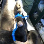
Mia is an older lab mix who had a progressively growing mass in the middle of her tarsus (between the ankle and the foot). It was not painful, but it was getting big enough to cause her family concern. She was brought to her regular vets office, a corporate practice, who wanted to refer her to a surgeon for the removal. Mia’s family came to see me for a second opinion.
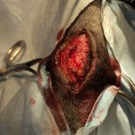
Skin mass of unknown origin. Raised, irregular, nodular mass about 5 cm widely adherent at the base to the underlying tissue. The difficulty in this surgery lay in the size of the mass, the width of the base, the lack of adequate tissue to closely easily, and the mass had no previous diagnostics done on it, therefore we were removing ti without the benefit of knowing what it was most likely to be.. Not knowing what it is makes the chances of complete excision more difficult. In many cases like these we are more accurately taking a biopsy with an incomplete excision, which in some cases really matters to long term prognosis and after surgery options.
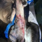
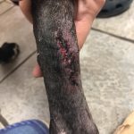
Mass removal via laser surgery. Mia was given pre-operative blood work. Surgery consisted of; i.v. fluids, laser, post op bandaging to protect the incision and manage tension at the site.
Mia did very well. Best of all her biopsy results were wonderful! (always biopsy an unknown mass!)
Here is her biopsy report:
DESCRIPTION:
This is a pedunculated mass which the dermis is expanded by
disorganized, abnormally enlarged hair follicles, sebaceous glands,
sheets of well-differentiated adipocytes, and thick mature brightly
eosinophilic collagen. Ruptured keratin and hair shafts are
surrounded by large numbers of neutrophils, macrophages, and plasma
cells supported by granulation tissue. The overlying epidermis is
markedly hyperplastic and hyperkeratotic, to ulcerated. The lesion
has been adequately excised.
MICROSCOPIC FINDINGS: FIBROADNEXAL DYSPLASIA WITH RUPTURE AND SEVERE
PYOGRANULOMATOUS FURUNCULOSIS, COMPLETE EXCISION
COMMENTS: Fibroadnexal dysplasia is a benign proliferation of adnexal
structures. These can also be known as fibroadnexal hamartoma or
fibroadnexal nevus. These are common in the dog and can become
evident at any age. Typical locations are along pressure points on
the limbs. Often adnexal structures in these lesions can become
disrupted and rupture, causing abundant inflammation. There is no
evidence of neoplasia in the sections examined.
Note; case submitted with owner permission


